Design and Fabrication of Micro Electrode Array (MEA) to evaluate functionality of neural cell network and cardiac cells
Planar microelectrode arrays have been used to characterize the electrophysiological properties of excitable tissues derived from both the brain and the heart. By using Signals we can detect the momentum of the action potential and minor oscillations. These electrodes are the connecting line between neurons and Electric circuits. There are two general classes of MEAs: implantable MEAs, used in vivo, and non-implantable MEAs, used in vitro.
Stimulating neurons and muscle cells causes ion currents to flow through the cell membrane, creating a potential difference between the inside and outside of the cell. Non-invasive connection between the electrode and the cell provides long-term recording and multi-week stimulation. By controlling the temperature and pH of the environment, electrical activities of the neurons cultured on these electrodes can be kept alive for more than a month.
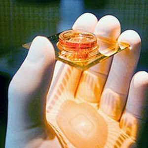
The MEA-Systems record, amplify, and analyze signals from biological samples in vitro. The data is analyzed by the included data acquisition software. MEA-Systems are used to record from brain or cardiac slices, neuronal or cardiac cultures, ex vivo retina, cell lines or stem cells.
System features:
- Stable and fully biocompatible surface for cell culture
- Non-invasive recording
- Long-term recording
- capability
- Good spatial distribution for getting a map of cell excitation patterns with arrays
- Fast results
- Easy to use compared to patch clamp method